


 Metatarsus Varus
 Metatarsus Varus
What is metatarsus Varus?
The Metatarsus Varus is an assemblage of bones in the central section of the foot. Each foot has five metatarsal bones, each conjugated to the phalanges of the toes.
Metatarsus primus varus refers to a condition where the metatarsal bones are spun toward the middle of the body. This causes an apparent defect, and both feet are frequently affected.
It’s a frequent congenital disorder in babies that are supposed to be caused by intra-uterine positioning that leads towards the aberrant adduction of the forefoot at the tarsometatarsal joint.
May be “flexible” (the foot can be uncurled to a degree by hand) or “non-flexible” (the foot cannot be uncurled by hand).
Occurs in about one of every 12 live births. Babies with Metatarsus Adductus are at augmented risk for developmental dysplasia of hipsterism.
In this deformity, also called the Pigeon Toed the frontal part of a child’s foot turns inward. There’s no given cause of metatarsus varus. It is a veritably common foot condition in children.
Most cases of “MA” resolve without treatment. Still, in over 14% of affected children, the disfigurement persists to adulthood [1, 2]. Numerous pediatricians regard metatarsus varus as a cosmetic issue of minor functional importance [3]. Still, it’s well demonstrated that unresolved metatarsus varus may need additional treatment [4-9].
Epidemiology
Metatarsus varus occurs in approximately 1 in every 12 births and has an equal frequency in males and females. This common malformation in babies is bilateral in approximately 60% of cases.
There is an increased incidence in late pregnancy, first pregnancies, twin pregnancies, and oligohydramnios. Associated conditions include DDH (15-20%) and torticollis. [10].
Although most cases of metatarsus varus resolve by the time of skeletal maturity, the persistence of the deformity has been associated with
the development of hallux valgus (HV). The incidence of MA in patients with HV was reported to be 21.6% to 29.5% [11].
What causes metatarsus varus?
Metatarsus varus is thought to be caused by the infant’s position inside the womb.
Risks may include that the baby’s bottom was pointed down in the womb (breech position) or the mother had a condition called oligohydramnios, in which she did not produce enough amniotic fluid.
There may also be a family history of the condition.
Metatarsus varus Diagnose
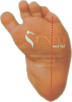
MA can be diagnosed through a physical exam. Telltale signs of this condition include the high arch and a visibly curved and separated big toe.
During the examination, the doctor will obtain a complete birth history of the child and ask if other family members were known to have metatarsus varus.
Diagnostic procedures are not usually necessary to evaluate metatarsus varus. However, X-rays (a diagnostic test that uses invisible electromagnetic energy beams to produce images of internal tissues, bones, and organs onto film) of the feet are often done in the case of nonflexible metatarsus varus.
An infant with this deformity has a high arch and the big toe has a wide separation from the second toe and deviates inward.
Flexible metatarsus varus is diagnosed if the heel and forefoot can be aligned with each other with gentle pressure on the forefoot while holding the heel steady.
This technique is known as passive manipulation. If the forefoot is more difficult to align with the heel, it is considered a nonflexible, or stiff foot.
Treatment
Specific treatment for metatarsus varus will be determined by your child’s doctor based on your child’s age, overall health, and medical history, the extent of the condition your child’s tolerance for specific medications, procedures, or therapies, expectations for the course of the condition and your opinion or preference.
In light of reports that 10–14% of metatarsus adducts cases persist to adulthood and some require surgical intervention, a more proactive approach is advocated by some authors for rigid, severe deformities [12].
The goal of treatment is to straighten the position of the forefoot and heel. Treatment options vary widely from watchful follow-up, passive stretching, bracing, serial casting, and, rarely, surgical correction [13].
Studies have shown that metatarsus varus may resolve spontaneously (without treatment) in the majority of affected children.
Metatarsus varus gets further complicated by the notion that early intervention, before 8 to 9 months of age, when spontaneous improvement is more likely, carries a better chance for deformity improvement [14].
When more severe, less flexible metatarsus varus is present, bracing or serial casting is often recommended with reported long-term success rates of approximately 90% [15, 16].
A doctor or nurse may instruct you on how to perform passive manipulation exercises on your child’s feet during diaper changes.
A change in sleeping positions may also be recommended. Suggestions may include side-lying positioning. In rare instances, the foot does not respond to the stretching program, long leg casts may be applied.
Serial casting series
Casts are used to help stretch the soft tissues of the forefoot. The plaster casts are changed every 1 to 2 weeks by your child’s pediatric orthopedist.
If the foot responds to casting, straight-last shoes may be prescribed to help hold the forefoot in place. Straight last shoes are made without a curve in the bottom of the shoe. For those infants with very rigid or severe metatarsus varus, surgery may be required to release the forefoot joints. Following surgery, casts are applied to hold the forefoot in place as it heals.
The UNFO Brace
The UNFO brace is a revolutionary treatment, invented by MD Izak Daizade to treat metatarsus varus, metatarsus adductus, pigeon-toed, and forefoot adduction deformities.
That kind of deformities among infants are called “FFA”, the UNFO brace is the perfect solution for newborns under 9 months only and not more than that.
The UNFO shoes can not treat an infant after they learnt to walk, that’s why the Brace is not an optional treatment for toddlers as well.
There are several outcome studies of metatarsus varus, with most patients presenting with benign and self-limiting metatarsus varus [17-20].
However, there are numerous reports of unresolved metatarsus varus treated surgically [21, 22]. Therefore, it would seem that before surgical intervention, most children with resistant metatarsus varus should be actively treated with casts or orthoses, potentially decreasing the need for subsequent surgical procedures.
A recent meta-analysis emphasized the importance of an accurate initial determination of the severity of metatarsus varus to be able to decide the appropriate treatment strategy. For mild, flexible metatarsus varus, observation is suggested.
More rigid metatarsus varus should be treated with casting/bracing. This study also stresses the lack of perspective, randomized clinical trials [14].
References:
- PONSETI, I.v., and J.R. BECKER, Congenital Metatarsus Adductus: The Results of Treatment. JBJS, 1966. 48(4): p. 702-711.
- Browne, R. and D. Paton, Anomalous insertion of the tibialis posterior tendon in congenital metatarsus varus. The Journal of bone and joint surgery. British volume, 1979. 61(1): p. 74-76.
- Rerucha, C.M., C. Dickison, and D. Baird, Lower extremity abnormalities in children. American family physician, 2017. 96(4): p. 226-233.
- Ghali, N., M. Abberton, and F. Silk, The management of metatarsus adductus et supinatus. The Journal of bone and joint surgery. British volume, 1984. 66(3): p. 376-380.
- BERMAN, A. and J.J. GARTLAND, Metatarsal osteotomy for the correction of adduction of the fore part of the foot in children. JBJS, 1971. 53(3): p. 498-506.
- Harley, B.D., et al., Abductory midfoot osteotomy procedure for metatarsus adductus. The Journal of foot and ankle surgery, 1995. 34(2): p. 153-162.
- HEYMAN, C.H., C.H. HERNDON, and J.M. STRONG, Mobilization of the tarsometatarsal and intermetatarsal joints for the correction of resistant adduction of the fore part of the foot in congenital club-foot or congenital metatarsus varus. JBJS, 1958. 40(2): p. 299-310.
- Mitchell, G., Abductor hallucis release in congenital metatarsus varus. International orthopaedics, 1980. 3(4): p. 299-304.
- Rodríguez, E.I.R., R.A. Reyes, and N.L. Marrero, New therapeutic approach of the congenital metatarsus varus and residual varus equine foot. Five-year study. Gaceta Médica Espirituana, 2014. 16(2).
- Loh, B., et al., Prevalence of metatarsus adductus in symptomatic hallux valgus and its influence on functional outcome. Foot & ankle international, 2015. 36(11): p. 1316-1321.
- Aiyer, A.A., et al., Prevalence of metatarsus adductus in patients undergoing hallux valgus surgery. Foot & ankle international, 2014. 35(12): p. 1292-1297.
- Mosca, V.S., The child’s foot: principles of management. 1998, LWW. p. 281,282.
- Bleck, E.E., Metatarsus adductus: classification and relationship to outcomes of treatment. Journal of pediatric orthopedics, 1983. 3(1): p. 2-9.
- Williams, C.M., A.M. James, and T. Tran, Metatarsus adductus: Development of a non‐surgical treatment pathway. Journal of paediatrics and child health, 2013. 49(9): p. E428-E433.
- Electricwala, A.J. and J.T. Electricwala, Congenital Quintus Valgus: An Extremely Rare Anomaly: A Case Report. JBJS case connector, 2017. 7(4): p. e75.
- Farsetti, P., S.L. Weinstein, and I.V. Ponseti, The long-term functional and radiographic outcomes of untreated and non-operatively treated metatarsus adductus. The Journal of bone and joint surgery. American volume, 1994. 76(2): p. 257-265.
- Reimann, I. and H. Werner, Congenital metatarsus varus: on the advantages of early treatment. Acta orthopaedica Scandinavica, 1975. 46(5): p. 857-863.
- Allen, W.D., D.S. Weiner, and P.M. Riley, The treatment of rigid metatarsus adductovarus with the use of a new hinged adjustable shoe orthosis. Foot & ankle, 1993. 14(8): p. 450-454.
- Berg, E.E., A reappraisal of metatarsus adductus and skewfoot. The Journal of bone and joint surgery. American volume, 1986. 68(8): p. 1185-1196.
- Galluzzo, A. and D. Hugar, Congenital metatarsus adductus: clinical evaluation and treatment. The Journal of foot surgery, 1979. 18(1): p. 16-22.
- Traversari, R., The metatarsal double osteotomy in the correction of hallux valgus with mini-invasive techniques. Interactive Surgery, 2007. 2(1): p. 38-43.
- Liabakh, A. and R. Rudenko, Modern status of the problem of hallux valgus surgery HALLUX VALGUS. Visnyk Ortopedii Travmatologii Protezuvannia, 2020(1 (104)): p. 77-84.


